https://medicine.ouhsc.edu/academic-departments Parent Page: Academic Departments id: 22853 Active Page: Subspecialty Areas of Studyid:22862

Subspecialty Areas of Study in Medical Physics

Diagnostic Physics

Diagnostic Physics

Includes radiographic and fluoroscopic equipment for routine and special x-ray procedures. Equipment is available for direct digitization of high-resolution fluoroscopically-generated images. This technique permits computer manipulation and analysis of the fluoroscopic images. Each clinical facility is provided with state-of-the-art Computed Tomography equipment. These units produce high-resolution images of internal human anatomy which are displayed as submillimeter distributions of tissue radiation attenuation coefficients. The Department operates a Magnetic Resonance Imager. Both 1.5 and 3 Tesla units produces anatomical images of the distribution of hydrogen/proton concentrations within tissue. This instrument makes available the most recent diagnostic procedures and offers the potential for considerable research as well.

MEDICAL ULTRASONOGRAPHY utilizes the principle of sonar to produce cross-sectional images of various organ and tissue interfaces in the body which are able to reflect high-frequency sound. Numerous units of various types are available for clinical and research us.

BI-PLANE - VASCULAR & INTERVENTIONAL SUITE utilizes high dose fluoroscopy for vascular imaging studies and repair, cardiac procedures and any procedure where time resolved x-ray imaging is required.

MAMMOGRAPHY is the special application of x-ray technology for detection of very small objects and low contrast objects in breast tissue. Mammography Quality Assurance Testing is one of several clinical experiences offered to first and second year graduate students.

Graduate students also receive hands on instruction from the medical physics faculty.

COMPUTED TOMOGRAPHY (CT) uses series of x-ray images to provide various reconstructed views (including 3D images) of patients for critical care investigations, surgical planning, interventional procedures and highly accurate anatomic modeling.

MAGNETIC RESONANCE IMAGING (MRI) uses powerful magnets and a series of high frequency pulses to obtain various reconstructed views. This type of imaging does not include the use of ionizing radiation and may be a viable alternative (as well as generally providing very high soft tissue contrast) for more vulnerable (radiation dose intolerant) populations.

NUCLEAR MEDICINE  deals with the applications of incorporated radioactive material, excluding brachytherapy, for diagnoses and treatment of diseases. Nuclear medicine procedures use radionuclide-labeled pharmaceuticals and scintillation crystal cameras to obtain the distribution pattern of the pharmaceuticals and to measure organ or tissue function, blood flow, or the presence of malignancy. The localization of the pharmaceuticals is used effectively for therapeutic radiation treatment as well as for palliative applications. Several of the recently developed scintillation camearas and computer systems are available in each of the nuclear medicine sections on campus and may be used for research purposes.  (PET/CT pictured on right and Cymbia SPEC/CT Gamma Camera shown below.)   

RADIATION BIOLOGY is the study of the effects of ionizing radiation on living things. Instrumentation is available to perform a variety of cellular and molecular techniques for the study of DNA damage and repair, changes in gene and protein expression, cell cycle regulation, and apoptosis. Students will have the opportunity to conduct radiobiology experiments to elucidate the mechanisms of radiation-induced cell killing, heat-radiosensitization, and effects of heat-shock and chemotherapeutic agents on cultured mammalian cells. In addition, students have access to several core facilities located on the campus.

Therapy Physics

Therapy Physics

Generally involves high radiation doses administered locally within the body to treat the cancer. The department uses modern equipment of radiation delivery and treatment planning.


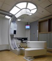
Mevion S250 Proton Therapy System
     – Pencil Beam Scanning, IMPT

     – First patient was treated

January 14th, 2019

     – 2nd center to treat patients in the U.S.

     – Upgraded from a double scattering system

     – RayStation treatment planning software

4 Linear Accelerators

     –1 TrueBeam Novalis STX  with HD MLCs
     –1 Varian Trilogy

     –2 Varian 2100EXs (one with OBI upgrade)

Varian Eclipse treatment planning software, BrainLab's iPlan for SBRT

     ~80 – 110 patients treated per day
   
HDR Brachytherapy

     –Elekta (Nucletron) microSelectron v3

     –Oncentra treatment planning system

     – ~10 – 15 patients per week: interstitials, cylinders, T&R & Venezia

LDR Brachytherapy

     –Eye Plaque: ~40-50 per year, I-125 with COMS/Eye Physics plaques

  • Varian BrachyVision / Eye Physics treatment planning system
    •Varian VariSeed LDR Planning System for Prostate Implant Radiotherapy
         –Prostate Seeds: ~ 2-3 per year

• Total Body/Skin Irradiation  

   – ~ 5-10 patients per year

Gamma Knife

     – Leksell Gamma Knife Perfexion

     – ~100 patients per year

     – Located in OU Medical Center