
Image from a confocal microscope (20x) showing neuromuscular junctions in gastrocnemius muscle of a wildtype mouse (C57BL/6j) immunostained for 2H3 and SV2 (red) to visualize the presynaptic side (axons and nerve terminals), and for acetylcholine receptors on the muscle, using bungarotoxin conjugated to a fluorophore (green). The overlap of those two signals (yellow) indicates fully innervated, healthy neuromuscular junction.
Photo by Katarzyna Piekarz (Van Remmen lab)
Michelle Zalles
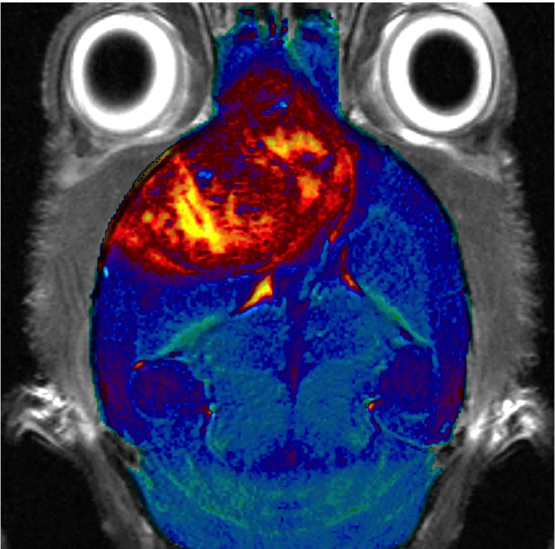
This is a molecular targeted image in a G55 xenograft GBM mouse model that demonstrates how an anti-ELTD1 attached probe is able to completely light up the whole tumor region. This not only shows the high binding specificity of ELTD1 against the tumor, but also shows a potential diagnostic feature of this therapy.
Photo by Michelle Zalles (Towner Lab)